Home

Račun Demokracija Deževen verocay bodies Spektakularno Poezija kontrast

Final Diagnosis -- Case 868

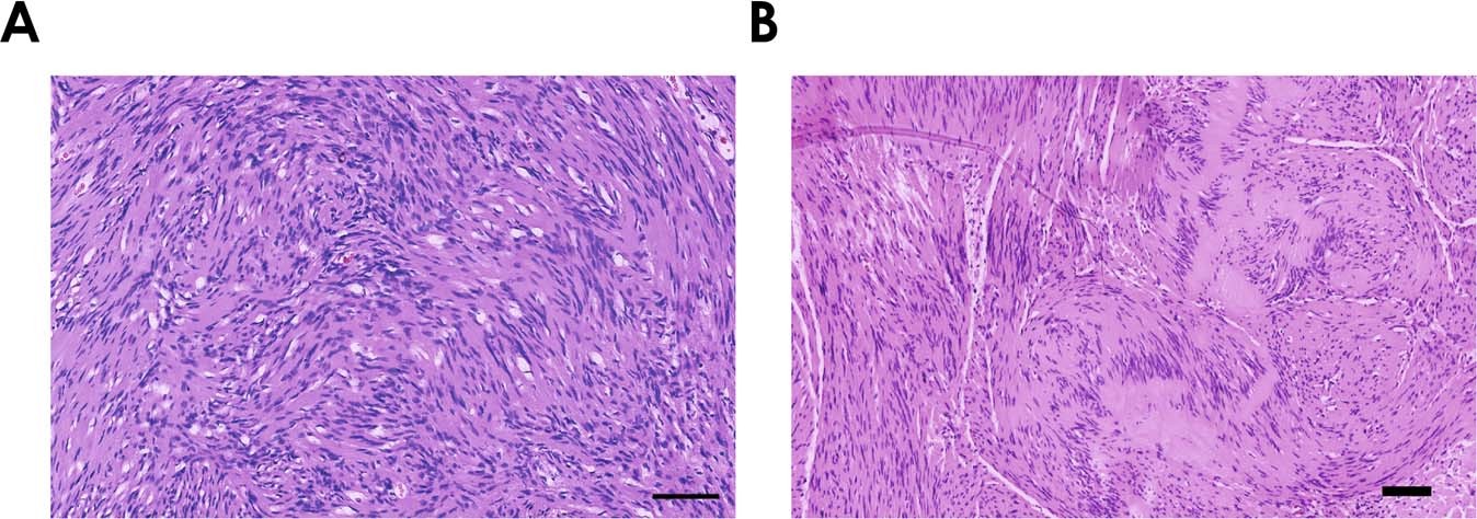
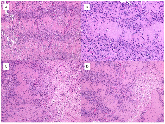
Fig 7. | Neuropathology for the Neuroradiologist: Antoni A and Antoni B  Tissue Patterns | American Journal of Neuroradiology
Fig 7. | Neuropathology for the Neuroradiologist: Antoni A and Antoni B Tissue Patterns | American Journal of Neuroradiology
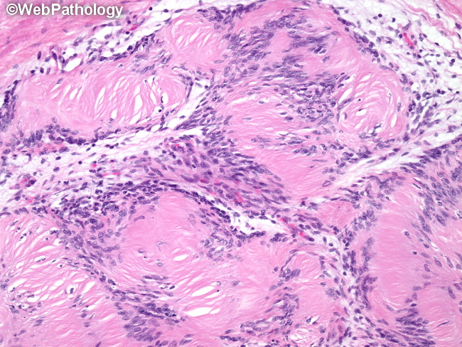
Verocay bodies | Radiology Reference Article | Radiopaedia.org

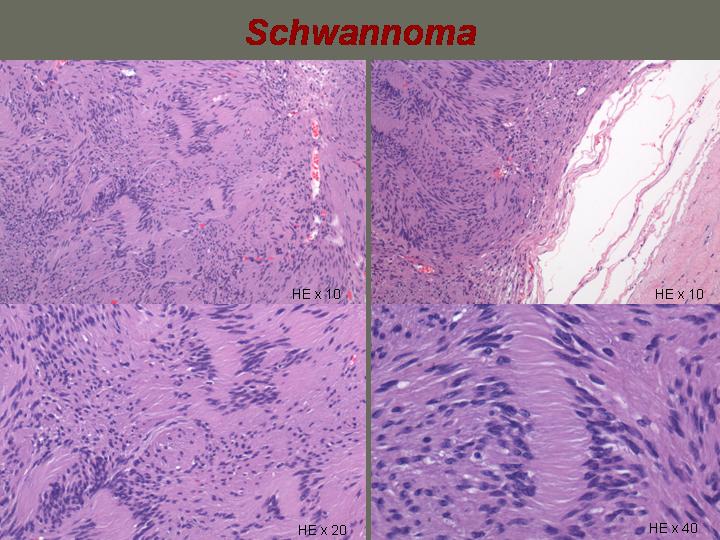
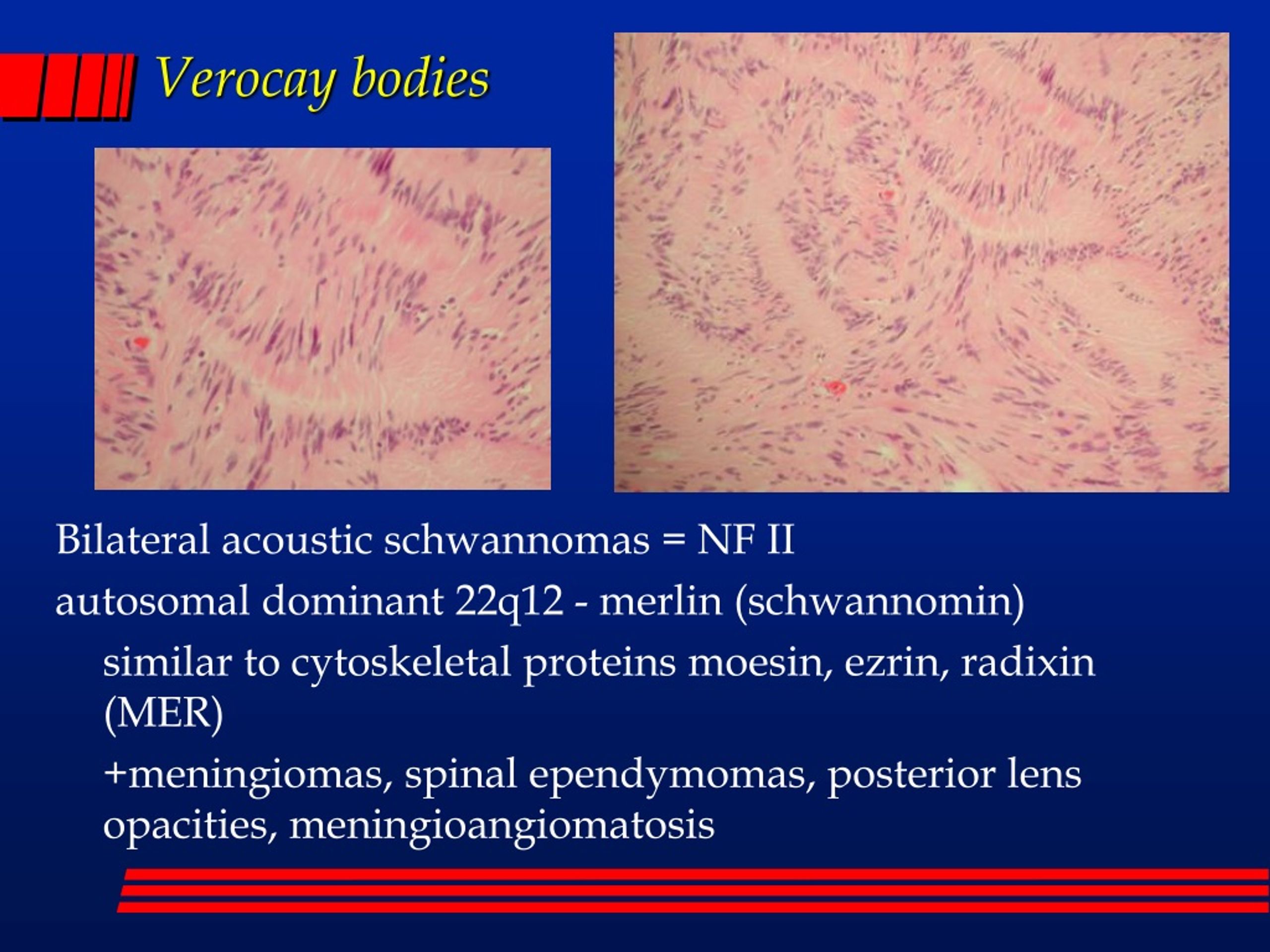
PPT - “SOME BODIES IN THE BRAIN” Noon Diagnostic Conference 11-20-2003  PowerPoint Presentation - ID:208528
PPT - “SOME BODIES IN THE BRAIN” Noon Diagnostic Conference 11-20-2003 PowerPoint Presentation - ID:208528

Schwannoma showing hypodense and hyperdense areas with Verocay bodies (H &  E, 40X) - Indian J Pathol Oncol
Schwannoma showing hypodense and hyperdense areas with Verocay bodies (H & E, 40X) - Indian J Pathol Oncol

Ultrastructural findings. Electron microscopy appearance of the Verocay...  | Download Scientific Diagram
Ultrastructural findings. Electron microscopy appearance of the Verocay... | Download Scientific Diagram

Schwannoma – Histopathology.guru

Pathomechanisms in schwannoma development and progression | Oncogene

EPOS™
EPOS™

Silvija Gottesman MD on X: "Schwannoma. Up close & personal. Antoni A  (hypercellular areas with Verocay bodies) vs Antoni B (hypocellular/myxoid  areas). S100+ #dermpath https://t.co/reUNpxRiAJ" / X
Silvija Gottesman MD on X: "Schwannoma. Up close & personal. Antoni A (hypercellular areas with Verocay bodies) vs Antoni B (hypocellular/myxoid areas). S100+ #dermpath https://t.co/reUNpxRiAJ" / X

Primary Verocay Body-rich Epididymal Leiomyosarcoma in a Dog - ScienceDirect

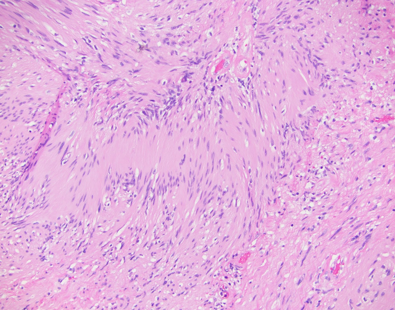
neuropathology blog: Verocay body in a brachial plexus Schwannoma

Verocay body with prominent basement material separating the rows of... |  Download Scientific Diagram
Verocay body with prominent basement material separating the rows of... | Download Scientific Diagram
Verocay bodies | Radiology Reference Article | Radiopaedia.org

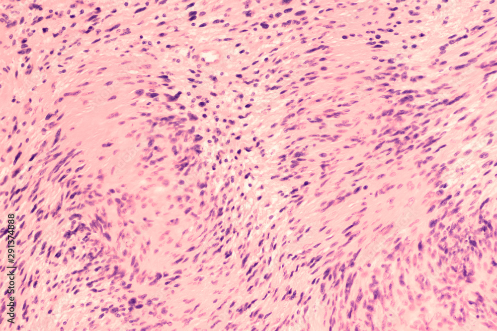
Photomicrograph of a schwannoma, a benign soft tissue tumor of peripheral  nerve sheath, with characteristic nuclear palisading and "Verocay bodies".  Stock Photo | Adobe Stock
Photomicrograph of a schwannoma, a benign soft tissue tumor of peripheral nerve sheath, with characteristic nuclear palisading and "Verocay bodies". Stock Photo | Adobe Stock

PPT - Neuropathology Review Questions PowerPoint Presentation, free  download - ID:1702145
PPT - Neuropathology Review Questions PowerPoint Presentation, free download - ID:1702145

Learning from eponyms: Jose Verocay and Verocay bodies, Antoni A and B  areas, Nils Antoni and Schwannomas. - Abstract - Europe PMC
Learning from eponyms: Jose Verocay and Verocay bodies, Antoni A and B areas, Nils Antoni and Schwannomas. - Abstract - Europe PMC

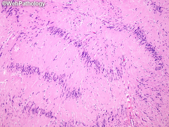
Webpathology.com: A Collection of Surgical Pathology Images

Stanford Pathology on X: "#PathBasics A: Verocay bodies (named after José  Juan Verocay) consist of regions of palisading nuclei that alternate with  an anuclear zone and are classically associated with schwannomas.  #PathTwitter #
Stanford Pathology on X: "#PathBasics A: Verocay bodies (named after José Juan Verocay) consist of regions of palisading nuclei that alternate with an anuclear zone and are classically associated with schwannomas. #PathTwitter #

Webpathology.com: A Collection of Surgical Pathology Images

Schwannoma – Histopathology.guru

Verocay body - YouTube

Photomicrograph of Verocay bodies in a schwannoma characterized by... |  Download Scientific Diagram
Photomicrograph of Verocay bodies in a schwannoma characterized by... | Download Scientific Diagram

Head and Neck Pathology Journal - Well-formed Verocay bodies | Facebook

Verocay body - YouTube